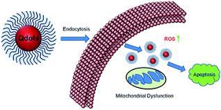
Quantum dots (QDs) are semiconductor nanocrystals that emit fluorescence on excitation with a light source.
They have excellent optical properties, including high brightness, resistance to photobleaching and tunable
wavelength. Recent developments in surface modification of QDs enable their potential application in cancer
imaging. QDs with near-infrared emission could be applied to sentinel lymph-node mapping to aid biopsy and
surgery. Conjugation of QDs with biomolecules, including peptides and antibodies, could be used to target
tumors in vivo. In this review, we summarize recent progress in developing QDs for cancer diagnosis and
treatment from a clinical standpoint and discuss future prospects of further improving QD technology to identify
metastatic cancer cells, quantitatively measure the level of specific molecular targets and guide targeted cancer
therapy by providing biodynamic markers for target inhibition.
Quantum Dots for Cancer Diagnosis and Therapy
Imaging is an important clinical modality used in determining appropriate cancer therapy. Current imaging
techniques, including x-ray, computed tomography, ultrasound, radionuclide imaging and MRI, have been used
widely for cancer screening and staging, determining the efficacy of cancer therapy and monitoring recurrence
(reviewed in).However, current imaging techniques have two major limitations. First, they do not have sufficient
sensitivity to detect small numbers of malignant cells in the primary or metastatic sites. Second, the imaging
techniques have not been developed to detect specific cancer cell-surface markers. In many instances, these
cell-surface markers might be targets for cancer therapy and might assist in the diagnosis and staging of cancer.
These limitations demand improvement in current imaging techniques and the development of new imaging
probes that are highly sensitive and biospecific. Quantum dot (QD) imaging probes, although still in the early
development stage, provide the potential to fulfill these requirements for in vivo cancer imaging.
Bioconjugation Quantum Dots are among the most promising items in the nanomedicine toolbox. These
nanocrystal fluorophores have several potential medical applications including nanodiagnostics, imaging,
targeted drug delivery, and photodynamic therapy. The diverse potential applications of Bioconjugation
Quantum Dots are attributed to their unique optical properties including broad-range excitation, size-tunable
narrow emission spectra, and high photostability.
The Drug delivery quantum dots nanocrystals fluoresce when excited by a light source, emitting bright colors
that can identify and track properties and processes in various biological applications. They have significant
advantages over traditional fluorophores as they can be predictably tuned according to their size, shape and
intrinsic solid-state properties. Their flexibility means it has applications in cell biology, drug discovery, cancer
research and other fields.
Millions of people die from cancer every year, especially from lung cancer. Even though no existing method can
defeat cancer, tumor therapies such as surgery, Quantum Dots Cancer Therapy tiny light-emitting particles on nanometer scale, are new type of fluorescent probes for molecular and cellular imaging. Compared with organic dyes and fluorescent proteins, Quantum Dots Cancer Therapy have unique optical and electronic properties in
cellular imaging: Wavelength-tunable emission, improved brightness of signal, resistance against
photobleaching, etc. Such preponderant optical properties were not realized until the QD-based probes are
equipped with war heads targeting tumor.
